Weekend Warriors and Heel Pain
Prepared by: Lisa Leff, PT BSc (PT), Cred MDT, Senior Manager, Quality and Outcomes, UHN Altum Health
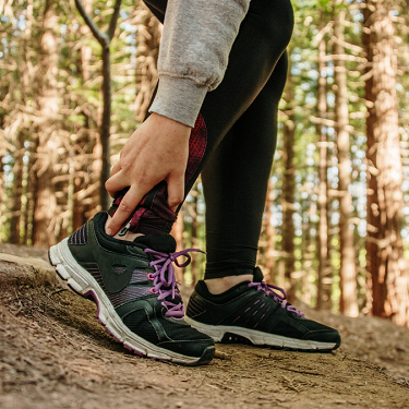
Many of us have experienced that annoying, sometimes excruciating pain in our heels that seems to come out of nowhere, that keeps us up at night and makes it difficult to stand and walk! Heel pain is a common condition that can be caused by a variety of factors, including overuse or injury, biomechanical issues, and underlying medical conditions. In fact, 10% of the population can develop what is called plantar fasciitis, one of a number of possible conditions causing heel pain.
Here’s some more possible reasons why you might develop sudden onset heel pain after activity:
Plantar fasciitis: This is a common cause of heel pain, and is caused by inflammation of the plantar fascia, a band of tissue that runs along the bottom of the foot.
Stress fractures: individuals who engage in a lot of physical activity, such as running or plyometric activities such as HIIT, may be at risk for stress fractures in the heel bone.
Tendonitis: The tendons in the heel can become inflamed and cause pain, particularly in people who engage in repetitive motions or overuse their heels.
Heel spurs: Small bony growths that develop on the heel bone can cause pain and inflammation, and may be the result of long-term stress on the heel.
Gout: Gout is a type of arthritis that can cause sudden, severe pain in the heel or other joints. It is caused by the buildup of uric acid crystals in the joint. There are a number of risk factors for gout including diet, weight, medical conditions such as diabetes, kidney disease, age, sex (men are three times more likely to get it) and family history of gout are some.
How does an Altum Health Physiotherapist approach a client complaining of “heel pain” incorporating evidence and best practices?
First and foremost, addressing this issue early and with a thorough evaluation is key. A Registered Physiotherapist is a good choice in terms of evaluating you for your heel pain. They will ask questions to understand fully your unique situation, complete a clinical evaluation and then determine what the specific cause of the heel pain is so that appropriate differential diagnosis can be provided. An evidence based treatment plan may be recommended depending on the specific cause of the heel pain. All with the goal to get you back on your feet!
The good news is that while heel pain can hurt a lot and impact our daily functioning, there is treatment including exercises to strengthen and stretch the muscles and tendons in the foot and ankle, as well as techniques to reduce inflammation and improve mobility and ultimately function, and many of these can be taught by the physiotherapist and then carried out at home by you as part of a home program.
Case Study:
Mr. Joe Smith is a 42-year-old Police Officer who presents at Altum clinic with left heel pain.
He reported that the pain started as he began a new fitness regime after a 10-year hiatus (aka weekend warrior!). After approximately 3 weeks of completing High Intensity Interval Training (HIIT) 30 minute sessions 3 x per week, he noticed a gradual onset of pain. It began as an intermittent, “nagging” ache, that gradually escalated. It is localised to the posterior plantar aspect of his heel with occasional radiation up his tendo-achilles and into the arch of his foot. He reports that it has been present for the past two (2) months.
He describes the pain as a constant dull ache, that is periodically sharp with activity. He rates the pain between 2-7 out of 10 on the numeric pain rating scale (NPRS), where 0 is no pain and 10 is the worst pain imagined. It is most severe in the morning when he first stands up, or after sitting for prolonged periods and improves as he walks around.
He also reported that the pain worsens with prolonged standing or walking, usually greater than 45 minutes and particularly on hard surfaces. It is relieved temporarily, with short periods of rest with rest. He is unsure if different footwear – for example his work shoes versus training shoes have any impact on his pain.
Past Medical History:
Mr. Smith endorses that he is generally fit and well and recently passed his annual medical and fitness assessment for the Police service “with flying colours”.
He denies any history of pulmonary or cardiovascular disease, high blood pressure, diabetes and any previous injuries to his left foot. He has never undergone any surgeries.
He does report a workplace injury to his right knee several years ago, after an altercation with a detainee, but reports that this fully resolved after a short course of physiotherapy.
He takes no medication on a regular basis, but reports that he tried OTC Advil and Tylenol for this pain with limited effect, so discontinued its’ use.
He is a non-smoker and reports alcohol consumption on special occasions only.
He has not undergone previous assessment or treatment prior to today.
Social History:
Police Officer for 20 years in a permanent full-time (40 hours per week) position. His role involves shift work where he can be on community patrol either in his car or walking. He is on his feet for a large portion of his day. There are some seated activities as he wraps up his day at the station. He is currently working full time hours and managing full time duties.
He lives with his wife and their 2 small children, ages 5 and 7. He enjoys being active with his children, but because of his pain has had to limit some of his activities, particularly at the end of his work day. He recently resumed physical exercise because his children are requiring less of his support now they are both in school.
Assessment:
On examination, Mr. Smith demonstrates bilateral pes planus; he reports that he has had “flat feet” for as long as he can remember. He walks with a slightly antalgic gait, with reduced stance phase and diminished push off on the left during the gait cycle, compared to the right. There is no obvious leg length discrepancy.
There is localised pain and acute tenderness to palpation in the heel region, specifically in the calcaneal insertion of the plantar fascia on the plantar aspect of the heel medially. He has minimally limited dorsiflexion with pain in the posterior of his left ankle/foot. He demonstrates difficulty performing the single leg heel raise test with increased pain on the eccentric action of lowering back to the ground.
Windlass test is positive, with heel pain produced with passive dorsiflexion of the toes, specifically at the end range MTP extension. This test is one of the differential tests used to diagnose plantar fasciitis.
All other objective and orthopaedic tests are noted within normal limits.
Analysis/Clinical Impression:
The clinical history, presentation upon evaluation, including risk factors, and objective assessment are indicative of diagnosis of acute plantar fasciitis.
The physiotherapist would provide Joe with the following explanation and education following her evaluation
“Plantar fasciitis is one of the most common causes of heel pain. It involves inflammation of a thick band of tissue that runs across the bottom of each foot and connects the heel bone to the toes (plantar fascia). The plantar fascia acts as a shock absorber and supports the arch of the foot during activity. Though it is generally poorly understood, there are several risk factors including pes planus or pes cavus and overactivity including prolonged standing or walking, as well as sudden bursts of forceful activity (such as would be seen in HIIT) that may cause micro tears to develop due to the repeated stretching and contracting of the tissue. Occasionally plantar fasciitis can be linked to the development of heel spurs as a result of longstanding tension on the plantar fascia. Up to 10% of the population can develop plantar fasciitis. Generally, it is a self-limiting condition with around 75% of cases resolving spontaneously between 3- 12 months.
A short course of conservative treatment, including Physiotherapy and Massage Therapy can be helpful during recovery.
Physiotherapy Intervention:
Following assessment, initial patient-directed treatment should be considered the first line of management.
With Joe, symptoms have been present for a period of 2 months. Normal tissue healing in an otherwise healthy individual may take anywhere from 6 weeks to 6 months.
Given that Joe has not engaged in any self management techniques it would be appropriate to educate him on the following
- Relative rest and activity modification – for example building short microbreaks into his work day where he can alternate long periods of standing and walking with seated breaks of 5-10 minutes. He should also consider not participating in further HIIT until the inflammation is resolved and he demonstrates sustained symptom improvement.
- Stretching – the plantar fascia is directly impacted by the length and tightness of the calf muscle complex – gastrocnemius and soleus, both of which merge to form the Achilles tendon inserting into the posterior heel, in close proximity to the plantar fascia origin. Research indicates a positive effect on plantar fasciitis symptoms with regular stretching of both gastrocnemius and soleus.
Joe is advised to perform bilateral stretches for both gastrocnemius and soleus every 2-3 hours, for up to 30 seconds each, repeating 3-5 times. He was advised to stop if stretches increased his pain overall but was educated that a temporary increase in symptoms (for a period of no more than 20 minutes) is a normal response at this stage in recovery. An internet link to specific, evidence-based stretches was shared to ensure he was able to reference appropriate technique upon leaving the clinic.
Cryotherapy as a therapeutic intervention. In this instance Joe reports his pain to be constant in nature, indicative of an inflammatory component. It may be appropriate for him to re-trial the use of an OTC NSAID, such as Advil, but benefit may also be gained with the application of localised cold therapy to his left heel. He was encouraged to use ice or a gel pack for 10-20 minutes several times per day as able. Warnings were given regarding skin protection and skin sensation to ensure no adverse effects such as skin/nerve damage.
- Heel cups/OTC orthotics – recommended for shock absorbing impact and also to provide relative rest to the plantar fascia by “shortening” the Achilles tendon thus reducing the load on the plantar fascia.
Joe was given a 2-week period to assess the impact of self management techniques on his overall pain and recovery. Should he be compliant but continue to see no improvements in his pain and symptoms, the following physiotherapeutic and massage interventions may also be considered:
– Taping – helps stabilize plantar fascia and provide support for the arch of the foot. Can provide temporary relief in offloading stress to the plantar fascia.
– Deep tissue/myofascial massage – can be performed by a Registered Physiotherapist or Registered Massage Therapist. Deep myofascial tissue massage can be effective for relieving pain and discomfort associated with plantar fasciitis. Deep myofascial massage of the plantar fascia, manually or with instrumentation, is thought to promote healing by increasing the blood flow to the injured fascia. The technique involves concentrated pressure being applied with slow strokes to the areas that are connected to the fascia. This includes the outer calf muscles, Achilles, heel and the sole of the foot.
Research has shown that a combination of deep tissue massage and routine stretching is more effective at pain management than ultrasound treatments and stretching.
One case study found that twice-weekly plantar fasciitis massage therapy helped to increase mobility and decrease pain so significantly that physically taxing activities caused little to no plantar pain for the subject, after having previously self-excluded from those activities due to pain levels. Deep tissue massage should only be considered in combination with other therapies noted above. Evidence also suggests that some patients may become reliant on the use of what is best described as a “passive modality” when short term pain relief is produced.
For long term effects, massage should be considered an adjunct therapy to promote pain relief and enable participation in an active and progressive therapy program.
– Electrotherapeutic interventions. There is limited clinical evidence to suggest the use of interventions such as ultrasound and laser produce any significant improvement in the acute phase of recovery. As such these types of passive modalities should be limited. Use of Extracorporeal shock wave therapy may be considered after at least 6 months of conservative treatment is trialled but deemed to be ineffective. Extracorporeal shock wave therapy is used to promote neovascularization to aid in healing degenerative tissue found in plantar fasciitis. Benefits of extracorporeal shock wave therapy are that it is non-invasive and offers the hope for a faster recovery time.
Post – acute phase recovery
Once recovery has begun, inflammation is settling and symptoms have improved, the progressive and focused reintroduction of activity is important in the remodelling and functional recovery phases of tissue healing, in order to prevent recurrence and reinjury. Given Joe had resumed exercise following a 10-year hiatus, and quickly developed symptoms, education is also provided on posture and mechanics during activity and exercise (including specific footwear/activity surface etc), consideration of a more graduated return and pacing for this weekend warrior as well as the need for long term stretch/cool down post activity.
If you are a weekend warrior and have any musculoskeletal injuries, aches and pains and would like to try our services you can book an appointment for physiotherapy or massage therapy at one of Altum’s Clinics click here …..
References:
1.De Garceau D, Dean D, Requejo SM, Thordarson DB. The association between diagnosis of plantar fasciitis and Windlass test results. Foot Ankle Int.2003;24:251-255.
2.Plantar Fasciitis Benjamin K. Buchanan; Donald Kushner https://www.ncbi.nlm.nih.gov/books/NBK431073/
3.Thomson CE, Crawford F, Murray GD. The effectiveness of extra corporeal shock wave therapy for plantar heel pain: a systematic review and meta-analysis. BMC Musculoskelet Disord. 2005;6:19.
4.Sun J, Gao F, Wang Y, Sun W, Jiang B, Li Z. Extracorporeal shock wave therapy is effective in treating chronic plantar fasciitis: A meta-analysis of RCTs. Medicine (Baltimore). 2017 Apr;96(15):e6621. doi: 10.1097/MD.0000000000006621. PMID: 28403111; PMCID: PMC5403108.
5.Juchli L. Effectiveness of Massage Including Proximal Trigger Point Release for Plantar Fasciitis: a Case Report. Int J Ther Massage Bodywork. 2021 Jun 1;14(2):22-29. PMID: 34079601; PMCID: PMC8133876.
6. Bernice Saban, Daniel Deutscher, Tomer Ziv, Deep massage to posterior calf muscles in combination with neural mobilization exercises as a treatment for heel pain: A pilot randomized clinical trial, Manual Therapy, Volume 19, Issue 2, 2014, Pages 102-108

